Advanced 3D Bioprinting for Cancer Research
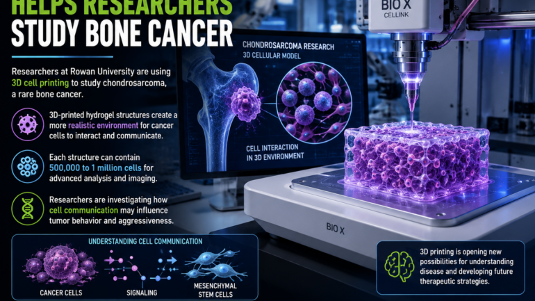
Researchers at the University of Mississippi Medical Center are leveraging advanced 3D bioprinting technology to create sophisticated three-dimensional models of tumors using living cells. This innovative approach is opening new avenues for studying cancer behavior and developing more effective treatments.
Bioprinting Technology
Unlike conventional 3D printers that use plastic filament to create mechanical parts, bioprinters deposit bioinks, which can contain living cells. These bioinks enable the construction of three-dimensional structures that closely mimic human tissue, providing a more realistic model for cancer research.
Miniature Tumor Models
The miniature tumors created through this process can indeed contain real cancer cells, arranged in a three-dimensional structure. These models capture the complexity of tumors more accurately than traditional two-dimensional cell cultures, offering a more comprehensive understanding of cancer behavior.
Advantages of 3D Models
Three-dimensional tumor models are particularly valuable because cancer cells do not grow in flat layers within the human body. They are surrounded by other cells, structural materials, and chemical signals. By replicating these interactions, researchers can better investigate how tumors grow and behave.
Testing Cancer Treatments
These models are also instrumental in testing experimental cancer treatments under controlled laboratory conditions. This allows researchers to identify promising drug candidates before advancing to more complex testing phases. However, it is important to note that results from these models do not directly predict how treatments will perform in patients.
Conclusion
The future of cancer research may well be built layer by layer through 3D bioprinting. This technology, with its ability to create intricate models using bioinks and living cells, holds significant potential for advancing our understanding of cancer and developing personalized treatment strategies.